Have your heard of the mythical character the Sandman? Legend has is that the Sandman drops fine grains on children’s eyes as they fall asleep. The sand prevents the eyes from reopening until morning so the children will remain in dreamland.
This magical man first appeared in a story by Hans Christian Andersen, where he was kind and told beautiful stories to kids as they slept. This nice version has made recent appearances in films like “Rise of the Guardians” and “The Santa Clause 2.” Other depictions of the Sandman are crueler and give him sinister motives.
Although you know the Sandman doesn’t exist, you might wish he would pay you a visit on nights when you struggle to fall asleep. Or, you may jokingly blame him for the goop you find in the corners of your eye every morning. But even without visits from the Sandman, your eyes have quite an interesting nightlife. Discover three facts about your eyes and sleep below.
1. Your Eyes Go Through a Carwash Every Night
Since the Sandman didn’t sprinkle sleep sand on your eyes, how did that goop get there? It accumulates as a result of your eyelids and tears combining to become a carwash for your eyes.
When you close your eyes for a long night’s rest, they stay firmly closed. Your tears wash over your eyes as they move around during different sleep phases. The tears pick up dust, old eye cells, mucus, bacteria, and any other small foreign substances in your eye. The closed eyelids guide the goop that ends up in the corners of your eye near your nose and along your lash lines.
The same cleaning process happens throughout the day when you blink. However, during those hours, the goop doesn’t get a chance to accumulate, so you don’t notice any gunk.
Typically, eye goop, which is properly called rheum, isn’t a sign of any eye condition. You can simply remove it with a warm, damp washcloth. But if you notice changes in your rheum, you may have an eye infection such as pink eye. Pay attention to rheum with an unusual color or consistency, and take note if your eyes produce more rheum than usual. Visit All About Eyes to rule out an infection if you experience these symptoms.
2. Blue Light Makes Your Eyes Want to Pull an All-Nighter
With the rise of smartphones and streaming video services, people spend many more hours a day interacting with electronics. And if you use your few precious hours before sleep relaxing in front of these devices, your sleep could be the victim.
Normally, our eyes use the light around us to figure out what time of day it is and cue our brains whether we should prepare for sleep. Light with wavelengths from the blue portion of the spectrum keeps us alert. Optimally, at night less blue light meets our field of vision so our brain begins releasing melatonin, a hormone that regulates our sleep cycle.
But devices with screens emit large amounts of blue light, as do LED lights. The cells in your eyes sense those wavelengths of light and pass on the message to the brain, disrupting the scheduled release of melatonin.
So far, there isn’t strong evidence that the blue light from smart devices does damage to your eyes. The sun itself can cause more harm. Still, sleep deprivation is linked to numerous health conditions, such as anxiety, depression, heart problems, weight gain, and type 2 diabetes.
If you want to avoid blue-light-induced sleep problems, try these tricks:
- Avoid devices that give off blue light at least 90 minutes before you hit the sack.
- Try apps or software that reduce the amount of blue light your devices give off.
- Uses glasses with lenses that block out blue light if you must use devices before bedtime.
Also, remember that blue light may only be part of the problem. Often the activity you engage in on your TV or smart device keeps your mind active, which can make falling asleep more difficult. Give yourself 30 minutes or so to clear your brain before bed so it can enter sleep mode.
3. Your Eyes Make Movies When You Dream
R.E.M. isn’t just an ’80s rock band—it’s also an acronym for what your eyes do while you dream. They make rapid eye movements. REM is one of several sleep stages, and it’s during this phase that our most vivid dreaming happens. Even if you don’t remember your dreams the next morning, your brain always dreams during REM sleep.
In recent years, scientists have discovered some fascinating facts about REM sleep. For example, they’ve learned that each eye movement creates a new image in your mind. Think of these as cells in a film strip or snapshots on your camera. Your eyes become the camera that pans around your dream world to take in the scenery.
Next time you prepare for a visit from the Sandman, remember that your eyes have a huge impact on your quality of sleep. Make some adjustments to how you treat your eyes if sleep has become elusive recently. And to keep your eyes in good condition no matter your sleep patterns, make an appointment at All About Eyes.
The human eye contains two different types of cells that interpret light and allow us to see. Rods allow us to see in low light. Because we only have one type of rod, they don’t convey color, which is why we generally don’t see color at night. We have three types of cones, however, which allow us to see colors ranging from red to violet in sufficient light.
Color blindness, more appropriately known as color vision deficiency, occurs when one or more of the cones has a defect, caused by a faulty gene on the X chromosome. Since men only have one X chromosome, they are much more likely than women to experience color blindness. For men, the chances of being colorblind are 1 out of 12, while for women the chance is 1 out of 200.
Types of Color Vision Deficiency
As mentioned above, humans have three types of cones: cones that perceive red light, cones that perceive green light, and cones that perceive blue light. One or more of these cones may have a defect, leading to poor perception of one or more colors. The different types of color blindness are described below.
Anomalous Trichromacy
With this first type of color blindness, each cone can still perceive color, but one does a slightly subpar job. People with this condition may be able to see the colors, just at a slightly reduced rate that makes it harder to distinguish different shades, or they may not be able to perceive the color at all.
Dichromacy
Unlike anomalous trichromacy, with dichromacy, one of the cones does not work at all. The individual can only see colors perceived by the two functional cones.
Monochromacy
With monochromacy, either only one cone is functional, or all three cones are faulty. In either case, the ability to perceive color is extremely limited, and many individuals only see in shades of black, gray, and white. To those with monochromacy, life is like a black-and-white movie.
What Colorblind People See
When those who see color correctly hear that another individual is colorblind, they often try to ascertain exactly what the world looks like to the other person. The answer can be complicated and changes according to what type of color vision deficiency the other person has one of the following:
- Protanomaly—faulty red cone. The colors red, green, brown, and orange all tend to have a similar hue. Instead of seeing a vivid distinction between red and green objects, both objects have a yellowish tint, with only a slight difference between the two colors. Blues and violets also appear similar, with little distinction.
- Deuteranomaly—faulty green cone. Like protanomaly, individuals have trouble distinguishing between red, green, brown and orange. Individuals with either type are referred to as red-green colorblind.
- Tritanomaly—faulty blue cone. This condition is the least common form of color vision deficiency. Individuals with tritanomaly see the world in shades of pink (standing in for orange, yellow, and red) and turquoise (standing in for blue, green, and violet).
- Protanopia—absent or dysfunctional red cone. The individual can’t detect any red light, so they see the world in shades of green. They can see yellow well but can mix up blues and purples, and dark red, dark orange, and dark green tend to look black or brown.
- Deuteranopia—absent or dysfunctional green cone. Like with protanopia, there is almost no difference between red and green colors. However, deuteranopes tend to see the world with a slightly more yellow hue than protanopes, who see more green.
- Tritanopia—absent or dysfunctional blue cone. This condition is the rarest form of dichromacy. Individuals will see gray instead of light blue, black instead of dark purple, blue instead of medium green, and red instead of orange. Pink is also more pronounced and yellow nearly impossible to see.
Those with color vision deficiency can live normal lives and don’t suffer too greatly because of their condition. Fashion can be a challenge, as a person with a red-green deficiency may not be able to tell whether a shirt is blue or purple or if a pair of socks is green or brown. To such a person, what they perceive as brown is the same as what they perceive as green.
Traffic lights may seem problematic since red and green are hard to differentiate, but colorblind people usually can drive safely based on the position of the light. A real issue is knowing when fruit is ripe or when meat is done cooking. To someone who can’t see subtle nuances between red, pink, and brown, a rare steak looks the same as a well-done steak.
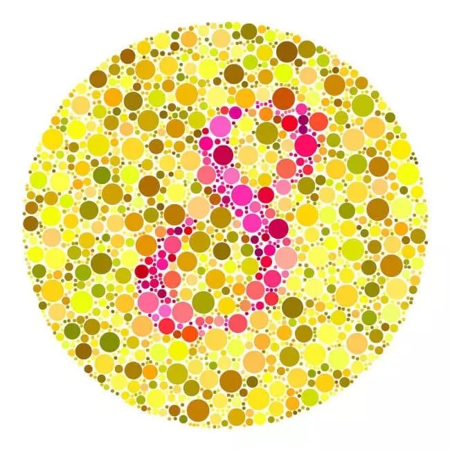
To get an idea of what the world looks like to a colorblind person, try out this vision simulator.
Possible Cures
Currently, no cure exists for genetic color blindness. However, a company called EnChroma has developed glasses that allow colorblind individuals to see wavelengths of light they aren’t able to detect naturally. Generally, people can now see red and pink where before they only saw green or brown. The glasses are mostly helpful for those with red-green color blindness, so they don’t work for everyone.
Scientists are also attempting to develop a treatment that involves an injection directly into an individual’s eye. The injection sends a virus into the eye to repair the defect in the cones. So far, the treatment has only been tested on animals, but the initial results show promise.
If you suspect that you or someone you know might be colorblind, visit your eye doctor to get tested. He or she will likely have some advice to keep your vision deficiency from interfering with any aspect of your life.
People notice eyes. We find eye contact important in daily conversation and human connection, and eyes have always fascinated human beings. So, when a pair of eyes have a unique or vivid feature, we immediately take note of it. Bright blue irises can be mesmerizing, and hazel eyes can easily draw attention.
Usually, iris color is what sets one pair of eyes apart from another. But, occasionally, we may notice something different about another’s pupils.
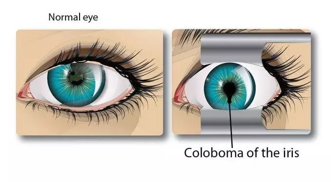
There is one optical condition that can result in keyhole-shaped or cat-like pupils from birth. These changes in appearance are attributed to coloboma, which can affect the lens, iris, retina, or optic nerve. The eye disorder can also affect the eyelids, but is more common in the eye itself.
Here, we discuss what coloboma is, how it’s developed, how it can affect the eye, and what kind of treatment or observation is recommended.
Condition
Essentially, the term coloboma is used to describe the condition where a portion of tissue is completely missing from the eye or eyelid. If a person has coloboma of the eyelid, part of the eyelid may be absent, and if they have coloboma of the iris, they may have irregularly shaped pupils.
The word is derived from the Greek word “koloboma,” which means “curtailed” or “defect.” It is a defect of the eye that is actually developed in the womb, so an individual would have the condition for their entire life.
It’s a fairly rare condition, and most studies can’t seem to agree just how rare because there could be numerous individuals that haven’t been diagnosed. According to the collection of studies, coloboma can affect 0.5 to 2.2 people out of every 10,000.
Development
During the fourth week of pregnancy, a baby’s eyes begin to develop. The eyes essentially grow from the brain, starting out as simple stalks. Eventually, a major portion of these stalks becomes the optic nerves, and the ends of these stalks become the eyes.
Underneath these stalks, there’s a seam that runs from end to end named the optic fissure. At five weeks in the womb, this optic fissure starts to close, starting at the center of the seam. In rare cases, the optic fissure doesn’t completely close at the ends, resulting in coloboma of the eye.
If the fissure doesn’t close near the front, the baby can have coloboma of the iris or lens, and if the fissure doesn’t close near the back, they can have coloboma of the optic nerve or retina. However, it is possible to have more than one of these areas affected.
Cases of coloboma can be completely isolated, which means it isn’t a feature in a more extensive condition. It can happen unexpectedly with no real origin, or it can be passed on to children from parents through either recessive or dominant genes. Coloboma can also be a part of another condition, such as fetal alcohol syndrome or CHARGE syndrome.
Effect on Vision
Most of the time, coloboma is identified by the unusual shape of the iris, and an individual may not experience any issues with their vision or eyes at all. However, coloboma of the retina or optic nerve can impair a person’s vision, and the severity of the impairment can range from mild to serious. Coloboma can also result in a sensitivity to light.
Also, there are cases where coloboma will affect the individual’s vision, but the iris and lens are completely untouched. This can make it a bit harder to identify, but not impossible to diagnose.
While an individual has coloboma from birth, the disorder can cause other eye conditions later in life, including cataracts, microphthalmia, glaucoma, or retinal detachment.
Treatment and Observation
When a baby is discovered to have a coloboma, either by the hospital staff at birth or a parent, it’s best to have an ophthalmologist take a look and run a series of tests. While the ophthalmologist may be able to learn more about the baby’s issue, they can’t determine if the baby’s vision is impaired until a later age.
It’s recommended that tests are done regularly as the baby grows to check for vision impairment or other eye conditions. Children with coloboma under seven years old should get semiannual checkups, and children older than seven should have annual checkups. If an issue should develop, the ophthalmologist can catch it early and treat it accordingly.
Currently, there’s no way to completely repair or cure this disorder, but if an individual with coloboma of the iris is dissatisfied with their appearance, they can get cosmetic contacts to cover up the iris. In some cases, they may even be able to surgically repair the iris.
Coloboma is a fairly rare condition, but people can contract all kinds of eye conditions. If you’re having trouble seeing or are experiencing any other kind of eye discomfort, visit a local eye doctor, such as those at All About Eyes. We can take a look at your eyes, determine the problem, and discuss possible solutions with you.

People of all ages recognized and admired the late David Bowie, whether it was due to a childhood appreciation for Labyrinth or a lifelong love for Bowie’s music. Many remember his eccentric outfits and makeup in his early career, and his unique personality and style have left a mark in history.
But one of Bowie’s most unique traits was, and still is, the difference between his eyes. At first glance, it appeared his eyes were two different colors, but upon closer inspection, his pupils were actually two different sizes.
This condition is actually called anisocoria, and it’s not as uncommon as you might think. But what exactly is anisocoria? And what causes it? Below, we’ll discuss what anisocoria is and how it develops, as well as how David Bowie came to contract this condition.
What is Anisocoria?
Anisocoria is a condition where the pupils are two different sizes. The word “anisocoria” literally translates into “not equal pupil condition.” However, while Bowie had a permanent case of anisocoria, the condition isn’t always lasting. And the difference in pupil size isn’t always as noticeable as Bowie’s; the difference between the two can be as little as 0.4 millimeters.
Surprisingly, anisocoria affects about 20% of the healthy population, and most of the time, the difference in pupil size is between 0.4 and 0.5 millimeters, but it can be up to a maximum of one millimeter. And Unequal pupil sizes can be caused by a variety of conditions.
What Causes Anisocoria?
Unequally sized pupils can actually be genetic. Children may be born with a difference in pupil size, which they may share with a parent or other direct relatives. If it’s a genetic condition, it’s not usually an issue.
Sometimes, aerosol medications or eye drops can cause anisocoria in a healthy individual. Again, this isn’t much of a concern, as long as the pupils return to normal after a reasonable amount of time.
However, anisocoria can be a result of serious trauma or conditions, such as:
- A brain abscess or tumor
- Intracranial hemorrhaging
- Brain swelling
- An aneurysm
- A stroke
- Inflamed membranes surrounding the brain
Anisocoria can also be caused by migraines or glaucoma. Should you experience a seizure, the pupils may temporarily differ in size afterwards.
When Does Anisocoria Warrant Medical Help?
Because anisocoria can be caused by both serious and minor conditions, it can be difficult to determine when it’s time to see a doctor for anisocoria. If you notice a difference in pupil size, get help, especially if the change is sudden, unexplained, or persistent. If you experience anisocoria after hitting your head or injuring your eye, you should see a doctor right away.
Should you develop anisocoria, you may sometimes experience:
- Headaches
- A sensitivity to light
- Nausea
- Pain in your eye
- Double or blurred vision
- Fever
- Vision loss
If a difference in pupil size is accompanied by any of the above symptoms, seek medical help immediately. Your anisocoria may be a result of a more serious issue.
How Did David Bowie Develop Anisocoria?
David Bowie had different pupil sizes for his entire career, and with so many conditions that result in anisocoria, you may wonder what exactly caused Bowie’s unique eye appearance. Was it a genetic development? Was it caused by a serious condition?
Actually, David Bowie’s anisocoria was caused by a simple fight over a girl. As a youth, Bowie had a good friend named George Underwood. The two of them had been friends since they were nine and remained close over the years. But when they were 15, they both became interested in the same girl, and the two had a bit of a disagreement.
Underwood ended up punching Bowie directly in the left eye. When Bowie retold the story, he said it wasn’t a necessarily hard punch, but it was suspected that Underwood’s fingernail had scratched Bowie’s eye and essentially paralyzed the iris.
He was immediately taken to the hospital and eventually had two different eye surgeries. But for the rest of his life, the pupil remained dilated to give him his signature look.
However, despite everything, Bowie and Underwood remained friends for the rest of David Bowie’s life. While the two played music together as teens, Underwood eventually moved on to commercial art. In later years, he helped design the album sleeves for a few of Bowie’s albums, including The Rise and Fall of Ziggy Stardust and the Spiders from Mars.
Later on, Bowie actually thanked Underwood for his eye condition, saying it gave him “a kind of mystique.”
David Bowie’s eye condition ended up giving him a unique appearance throughout his career. But if you experience any unusual eye conditions or vision problems, be sure to see an eye doctor as soon as possible. An eye doctor can take a look at your eyes and determine the best course of action. If your prescription is outdated, he or she can also check your vision and renew your prescription.

When you have an autoimmune disease, your immune system damages healthy cells, including the ones in your eyes. Sometimes, the effect on your eyes can be so pronounced that an eye doctor can be the first one to suspect you have an autoimmune disorder.
Maybe you’ve been diagnosed with an autoimmune disorder and are wondering how it effects your eyes. Maybe you’ve noticed recurring eye problems and think a disorder might be to blame. In either case, understanding the relationship between autoimmune disease and eye health can help you know what to do to take care of your eyes.
1. Rheumatoid Arthritis
With rheumatoid arthritis, your immune system primarily attacks the lining of your joints, beginning with the joints in your fingers and toes. In response, the lining becomes inflamed, pushing down on and damaging the bone and cartilage it’s supposed to protect. The disorder can affect other parts of your body as well, including your eyes.
Dry eyes caused by the immune system attacking the tear ducts is the most common eye-related symptom associated with rheumatoid arthritis, but in some cases, the arthritis can directly attack the sclera, or white part, of the eye. The resulting inflammation (which is called “scleritis”) makes your eyes red, painful, and sensitive to light, and it can destroy your eye tissues if not treated.
An ophthalmologist can prescribe artificial tears to deal with the dry eye or anti-inflammatory drops to treat the scleritis.
2. Lupus
The immune system of those who suffer from lupus indiscriminately attacks any body tissues, including organs, joints, blood cells, and skin. Therefore, the symptoms associated with lupus are broad. If you have lupus, you’ll experience cycles of remission and flare-ups, and with each flare, different symptoms may present themselves.
Lupus can harm the eyes in a variety of ways, including by inflaming the eye tissues themselves, damaging the nerves that control movement and vision, and damaging the skin of the eyelids.
The most common eye issues experienced by people with lupus, however, are changes to the blood vessels in the retina. When the immune system attacks the circulatory system, not enough blood reaches the retinal blood vessels. The eye tries to fix the problem but simply creates more blood vessels, which also don’t have access to blood and so only restrict your vision further.
The primary method for dealing with retinal vasculitis is to treat the lupus itself. Lupus currently doesn’t have a cure, but its symptoms can be managed. Your doctor will likely prescribe anti-inflammatory medication to help restore blood flow.
If your optometrist diagnoses you with retinal vasculitis and you don’t have any known autoimmune disorder, talk to your general doctor about that possibility. Retinal vasculitis is associated with many autoimmune disorders and rarely presents on its own.
3. Psoriasis
Psoriasis causes an extreme buildup of skin cells on the surface of the skin. While skin cells generally have a month-long life cycle, those with psoriasis experience new cell growth every few days. This leads to patches of red, dry skin covered in scales. Environmental and lifestyle factors, such as stress, cold weather, skin injuries, and certain medications may cause the disorder to flare.
Psoriasis affects the skin all over your body, including your eyelids. You may develop red, scaly areas that cause pain when you open and close your eyes, or the skin may grow dry and crack. Since the skin around your eyes is so delicate, you’ll need to talk to your doctor about treatment options right away.
To treat the psoriasis on your eyes, your eye doctor might suggest using corticosteroids or eczema medication. You will need to be careful when applying any ointment or creams—they can cause damage if they come in contact with the eye itself.
4. Multiple Sclerosis
This autoimmune disorder specifically targets the brain and nervous system. The immune system attacks the nerves, which try to repair themselves with scar tissue. However, the scar tissue interrupts the signals traveling through the central nervous system, causing numbness, muscle weakness, lack of body control, pain, and reduced cognitive function.
In addition to all those symptoms, you may also suffer from vision problems. Vision problems may actually be one of the first signs that you have MS, which is one reason why regular eye exams are so important. A common initial indicator of MS is an inflammation of the optic nerve, known as optic neuritis, which restricts your vision by either blurring it or limiting it completely.
Other eye issues associated with MS include uncontrolled eye movements and double vision, due to weak muscles preventing your eyes from coordinating with each other. In every case, the symptoms usually resolve themselves during the course of general MS treatment, but your eye doctor might recommend corticosteroids as well.
Autoimmune disorders impact many aspects of your life, including your vision, but there are ways to overcome these eye problems and retain your sight. Make an appointment with All About Eyes to receive a professional examination and learn how to take care of your eyes.

As far as eyesight in the animal kingdom goes, humans see quite well. We can see stars glowing from millions of light-years away, we can see a wide range of colors, and we can see clearly for miles if our sight isn’t obstructed.
Of course, some people don’t see as well as others. Eyesight is measured relative to what other humans see, so if your vision is 20/20, you see at 20 feet what others with unimpaired vision see at 20 feet. If your eyesight is 20/50, you can see at 20 feet what a person with perfect vision sees at 50 feet.
With vision correction, we can achieve “perfect” eyesight for humans, but although our sight is pretty incredible and probably the sense we rely on the most, our vision pales in comparison to that of some other animals.
While it’s nearly impossible to definitively rank which animals have the best eyes, since vision covers a wide range of attributes, we can see how the superior eyesight of certain animals compares to human eyesight. Each animal in this list, like humans, relies heavily on its eyesight for survival.
Eagles
All birds of prey have excellent long-distance vision, but eagles stand out. They can see clearly about eight times as far as humans can, allowing them to spot and focus in on a rabbit or other animal at a distance of about two miles. While we can see a candle flame at that distance, a small animal camouflaged in its surroundings would be hidden from us.
Eagles can also quickly shift focus, allowing them to essentially “zoom” in on their prey. They also can see a wider range of colors than we can, allowing them to differentiate small changes in coloration in their prey, as well as see UV light.
As far as daytime vision goes, eagles, hawks, and falcons reign supreme. However, they don’t do as well during the night.
Owls
These nighttime predators take the torch from eagles as soon as the sun goes down. Unlike many birds with eyes that sit at an angle, owl eyes face directly forward, giving them incredible binocular vision. Although their large eyes can’t move or roll like human eyes can, owls can move their heads nearly all the way around, allowing them to have a 270 degree range of vision without moving their bodies.
Their large, tube-shaped eyes contain many more rods than human eyes, which allow them to be more sensitive to light. Their irises widen to allow more light to reach their retina at night. Because the iris adjusts, owls can also see during the day (unlike other nocturnal animals that can only see well at night), but their vision is slightly blurry and they cannot see colors well.
Owls and other animals with excellent night vision have a reflective surface behind their retina known as the tapetum lucidum. This thin layer allows light to reflect back into the animal’s eye after it’s already passed through, giving the animal two chances to collect an adequate amount of light.
Humans, on the other hand, don’t have this trait, so our day vision is vastly superior to our night vision.
Mantis Shrimp
Eagles may be able to spot a rabbit from the sky, but mantis shrimp might have the most complex eyes in the entire animal kingdom.
For example, humans have three types of cones in our eyes, allowing us to see the colors red through violet. Mantis shrimp, on the other hand, have 16 types of cones. While scientific research shows this doesn’t necessarily mean mantis shrimp perceive minute differences in color, or see colors we can’t imagine, it does indicate that mantis shrimp have an advanced color recognition system.
The set-up of their eyes allows the shrimp to move the eyes independent of each other without compromising vision (much like a chameleon). They therefore have a wide field of vision, and their color receptors allow them to pick up on small changes in color almost instantaneously. This can help them find prey and mates, and avoid predators.
Sheep and Goats
When considering the incredible vision many animals possess, you may not think to put grazing animals on the list. However, if you look closely at a goat’s eyes, you’ll notice that their pupils are shaped like horizontal lines.
Research has found that pupil shape and eye orientation depend highly on the activities undertaken by different classes of animals. Tall daytime hunters generally have forward-facing eyes with round pupils, while small animals like cats, snakes, and foxes that hunt during the day and night have vertical slit pupils that help them with depth perception as well as night vision.
Prey animals, like deer, horses, and elk, who spend their time grazing out in the open and must be constantly on alert, have wide and narrow horizontal pupils, which gives them a wider field of vision than nearly any other animal. When they lower their heads, their eyes also rotate so they have a constant view of the area around them.
While humans don’t have superb night vision, the ability to see UV light, or vision that extends beyond 180 degrees, we do have the perfect vision to suit our lifestyle needs. We can also improve our eyesight if we experience impaired vision, unlike any other animal.
If you need vision correction, come to All About Eyes to get an examination from one of our optometrists and learn about the glasses and contact options we offer.